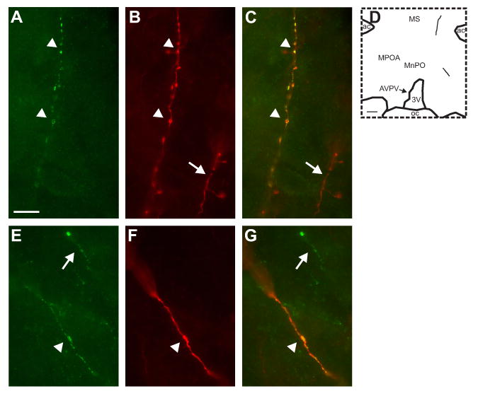
Figure 9.

High magnification photomicrographs of NKB (A and E, green) and BDA (B and F; red) immunofluorescence. The color-combined images (C and G) show that these NKB-immunoreactive axons are anterogradely labeled with BDA (yellow). The map in D shows the location of these dual-labeled axons. The top photomicrographs illustrate a thin, beaded anterogradely-labeled NKB-ir axon extending to the lateral ventral septal region (A, B and C, arrowheads). The bottom photomicrographs illustrate a dual-labeled axon in the medial preoptic region that is thicker and displays few varicosities (E, F, and G, arrowheads). The arrows show single-labeled fibers that are immunoreactive for either BDA (B and C) or NKB (E and G). Scale bar in A = 10 μm (applies to A-C and E-F). Scale bar in D = 200 μm.
