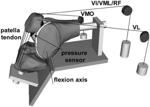
Figure 1.

Computational model of a flexed knee, embedded in a graphical representation of the in vitro testing frame. The loading cables used to apply forces through the quadriceps muscles are represented, along with representations of the pulleys and weights. The line segments representing the patella tendon are also shown, along with the sensor position within the patellofemoral joint and the tibiofemoral flexion axis.
