
Figure 6. Responsive and nonresponsive neurons show no difference in coherence with cortical slow rhythms.
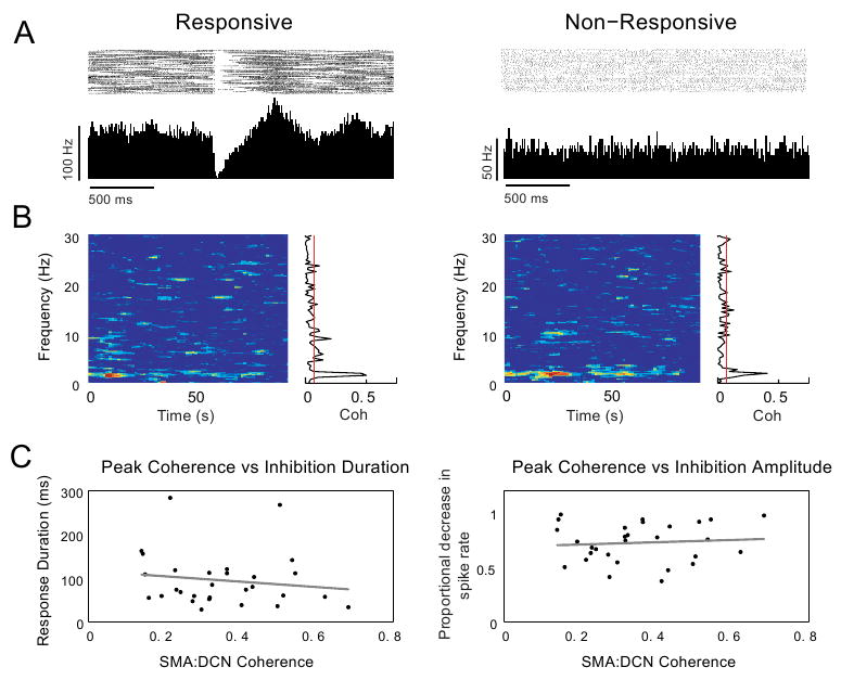
A-B: Coherence with cortical slow rhythms was compared between DCN neurons responsive to air puff stimulation of the lip and those that were non-responsive. In this example, a responsive and non-responsive neuron (peri-stimulus histograms and raster plots during stimulus-evoked activity shown in A) share the same coherence peak and profile (coherence during spontaneous activity of the same respective neurons shown in B; cf. left panels in A and B (responsive neuron) with right panels in A and B (nonresponsive neuron) (n = 1 animal). C: Coherence with cortical slow rhythms also does not predict the duration or amplitude of DCN neuron response components. A linear regression analysis showed no correlation between the response durations or amplitudes of the short-latency excitation (p > 0.05, n = 34), inhibition (p > 0.05, n = 57) or long-latency excitation (p > 0.05, n = 35) components to the peak frequency of coherence in responsive neurons. Scatter plots for inhibition duration and amplitude vs. peak coherences are shown in C.