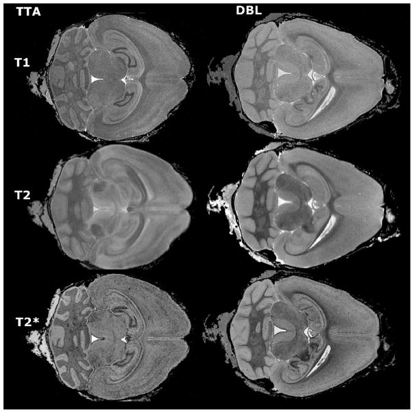
Figure 1.
Different imaging spectrums offer complementary information about brain structure. Multispectral images were acquired for both APP/TTA double transgenic mice (DBL) and TTA single transgenic controls (TTA). T1-weighted images provided the best structural definition and were used for segmentation (top row), T2-weighted images generated the best signal to noise ratio for detecting large amyloid plaques (middle row), while T2* (GRASS) images offered the best resolution for smaller deposits (bottom row).

