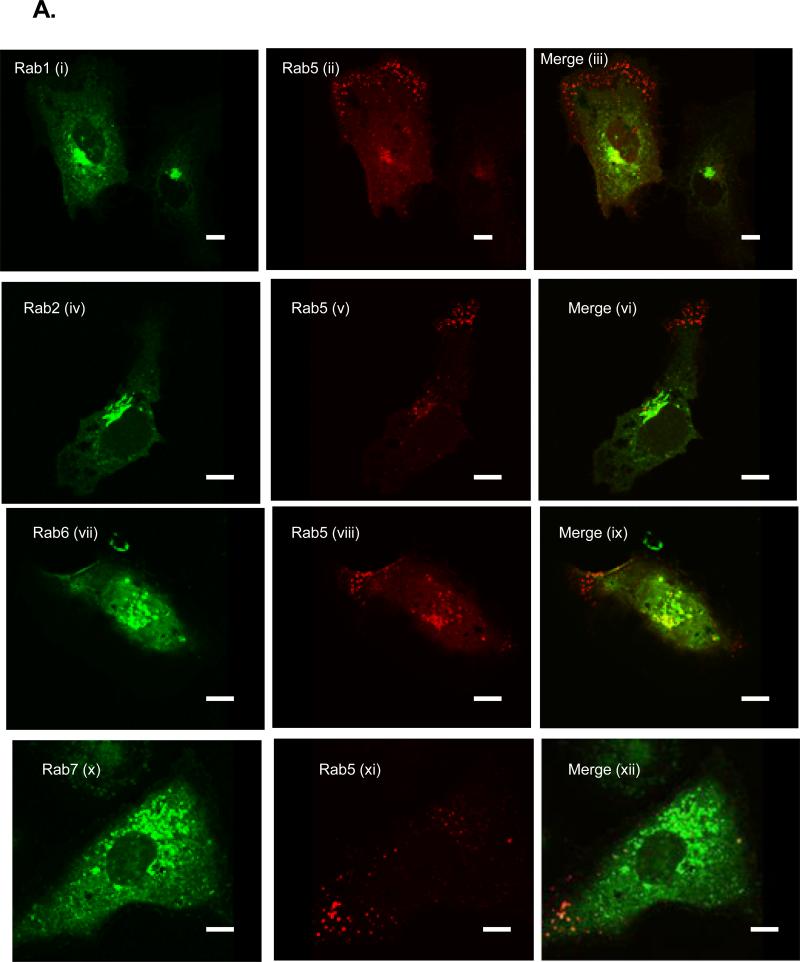
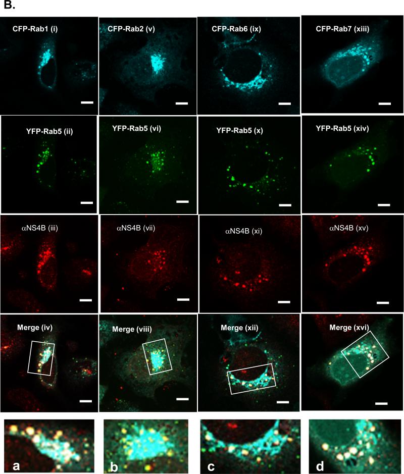
FIG.5.
A. Huh7 cells were co-transfected with DNA constructs expressing YFP-Rab5 and CFP-Rab1 (i-iii), 2 (iv-vi), 6 (vii-ix) or 7 (x-xii) proteins. At 24 h post-transfection, the cells were processed and pictures were taken with a confocal microscope. Note that the CFP-expressing Rab proteins were pseudo-colored green whereas Rab5 is red. Also notice the lack of a significant merging of Rab5 fluorescence with the other Rab proteins. B. C5B cells were transfected with DNA constructs expressing YFP-Rab5 and CFP-Rab1 (i-iv; a), 2 (v-viii; b), 6 (ix-xii; c) or 7 (xiii-xvi; d) proteins. At 24 h post-transfection, the cells were stained with NS4B-specific antibody and samples were examined as in (B). CFP-fused Rabs are blue, Rab5 is green and NS4B is red. Merging of the three colors results in white fluorescence. Magnified areas are indicated by rectangles. C. Quantitation results are from at least five cells co-expressing Rab proteins and NS4B. Notice the colocalization of Rab5 and 7 with NS4B in C5B cells (xiii-xvi; d) as displayed by the Pearson colocalization results in (C) and the white foci in (B). There is weak merging of Rab5 & 6 (ix-xii; c) or Rab5 & 1/2 (i-viii; a & b) with NS4B foci. Bars = 10 μm.


