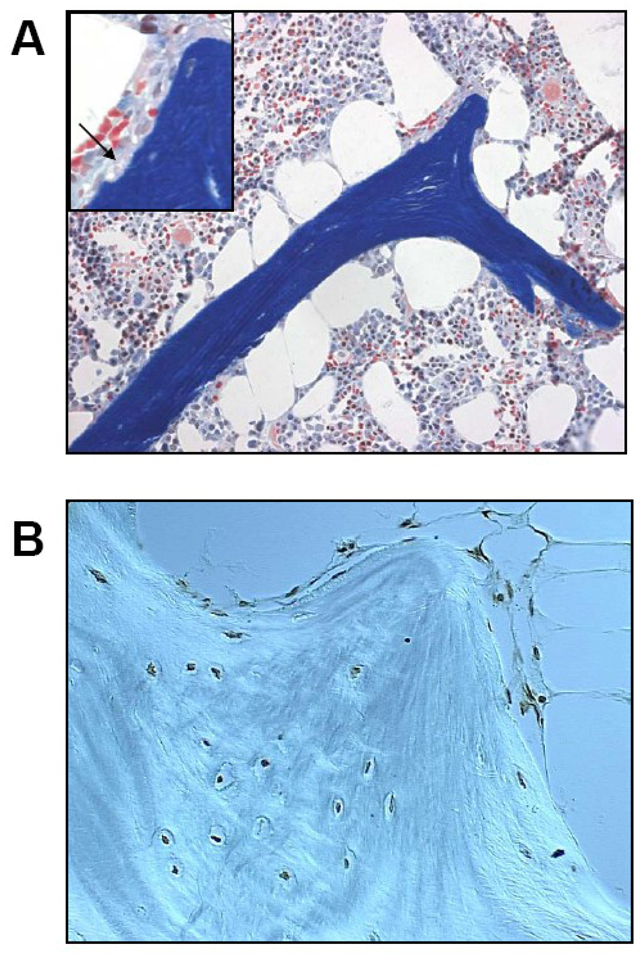
Figure 1. Glucocorticoid-induced osteoporosis.
(A) After long-term prednisone treatment in humans, cancellous bone tissue often shows an excessive number of enlarged adipocytes and atrophic, disconnected trabeculae without osteoblasts. The insert shows the accumulation of erosion cavities devoid of osteoclasts (arrow), measured as the reversal perimeter, which indicate delayed or defective bone formation rather than excessive bone resorption. The red cells in the bone marrow are hematopoietic cells and should not be mistaken for osteoclasts (Modified Masson staining, 200x with the insert at 400x). (B) In this specimen obtained from an osteonecrotic hip, virtually all cancellous osteocytes and lining cells are apoptotic as revealed by the dark brown stain (apoptosis staining was done by in-situ end-labeling or ISEL, 630x).

