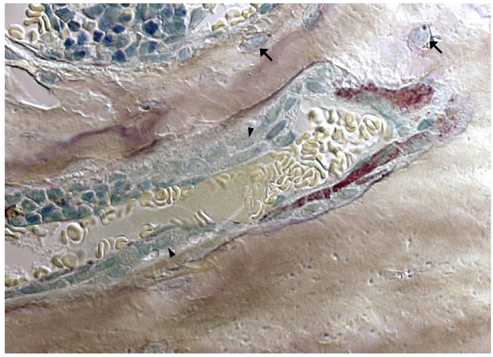
Figure 3. Each new basic remodeling unit of bone has a nearby blood vessel.
Murine osteoclasts with discrete tartrate-resistant acid phosphatase-positive red granules burrow deep into vertebral cancellous bone. Pale yellow erythrocytes are seen in the adjacent blood vessel that served as the conduit through which the osteoclast precursors were delivered to the remodeling site. Trailing behind the osteoclasts, teal-colored osteoblasts forming new bone are bringing up the rear (arrowheads). Osteocytes (arrows) are seen buried in the mineralized bone matrix. Methyl green and tartrate-resistant acid phosphatase-staining of undecalcified bone viewed with Nomarski differential interference contrast microscopy (x630).

