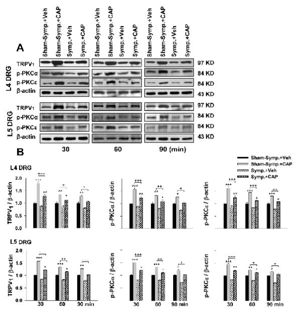
Figure 3.
Western blot analyses of the evoked changes in the relative density of TRPV1, p-PKCα and p-PKCε in L4 and L5 DRG tissue following CAP injection in sham-sympathectomized and sympathectomized rats at 30, 60 and 90 min after unilateral injection of CAP. Immunoreactivity of TRPV1, p-PKCα and p-PKCε was normalized to β-actin. A. Representative Western blots for TRPV1, p-PKCα and p-PKCε to CAP injection at 30, 60 and 90 min after injection in the sham-sympathectomized and sympathectomized rats. B. Grouped data of changes in these molecules. * P<0.05, ** P<0.01, *** P<0.001, compared with vehicle injection at the same time point in the group of sham-sympathectomy or sympathectomy. +, P<0.05, ++ P<0.01, +++ P<0.001, compared with the CAP-induced effects under sham-sympathectomized conditions.

