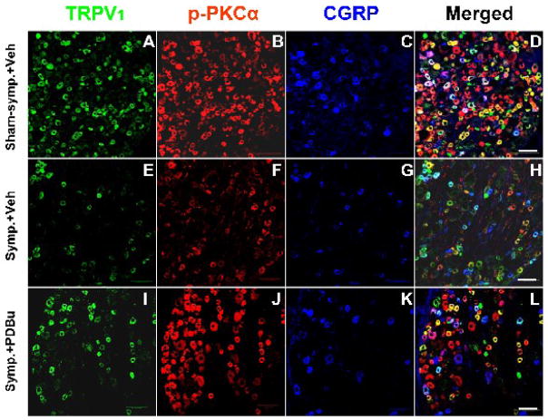
Figure 7.
Confocal immunofluorescence images showing changes in the CAP-evoked single labeling of TRPV1, p-PKCα, and CGRP and double or triple labeling of these molecules in neurons of the L4 DRG on the side ipsilateral to CAP injection after sympathectomy (E–H, Symp.+Veh) compared to the CAP-evoked changes in the sham-sympathectomized (A–D, Sham-symp.+Veh) and the effects of PKC activation by pretreatment with PDBu (I–L, Symp.+PDBu). Data were sampled at 30 min after CAP injection. Scale bar=100 μm.

