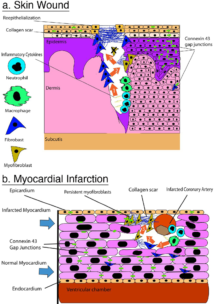
Figure 1.
A Comparison of scar formation in the skin and heart. Sequence of orange arrows in A and B convey sequential events in the healing process of each tissue. A) Skin wound, (Arrow 1) After hemostasis neutrophils and macrophages release proinflammatory cytokines for the destruction of dead tissue and invading organisms. (Arrow 2) Fibroblasts are recruited to the wound area and begin to lay down collagen to form a new dermal composite: granulation tissue. Myofibroblasts expressing α-SMA appear either from epithelial-mesenchymal transformation or circulating fibrocytes and continue to upregulate collagen expression. (Arrow 3) Collagen weave fully encloses the wound area and keratinocytes re-epithelialize from the periphery of the collagen rich scar. Myofibroblasts undergo apoptosis and are no longer detected. Notice Cx43 expression is down regulated in the epidermis proximal to the wound edge B) Heart Scarring post-MI (Arrow 1) Infarcted coronary artery results in death of local myocytes and recruitment of neutrophils and macrophages to the site of injury where they release pro-inflammatory cytokines. (Arrow 2) Fibroblasts and myofibroblasts are recruited from the surrounding myocardium and begin to lay down collagen. Note that Cx43 is down regulated in the infarct border zone and no longer localized at the end-to-end abutments of the myocytes. (Arrow 3) Myofibroblasts expressing Cx43 and forming intercellular junctions with adjacent myofibroblasts persist in the dense collagen scar.

