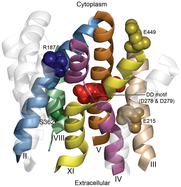
Fig. 8. Functional residues mapped on the structural architecture of NHA2.
The cytoplasmic side is upwards. The model-structure of NHA2 is shown as transparent ribbons, whereas helices implicated in the function mechanism are marked. Helices 9 and 12, along with the extramembrane loops, were omitted for clarity. Specific residues suggested to participate in transport, illustrated in Fig. 7C, are shown as spheres and highlighted.

