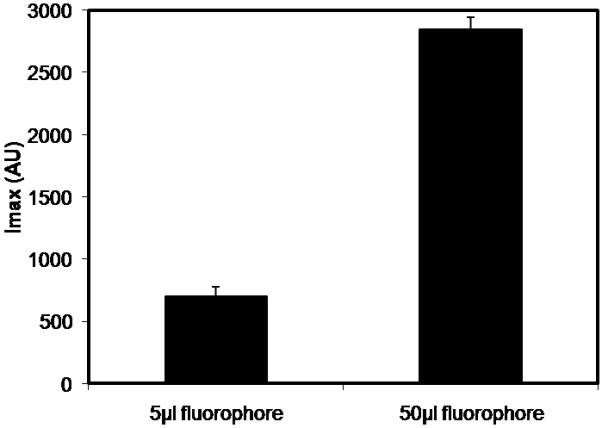
Fig. 6.

Comparison of maximum intensity values at the bead surface under different IL-6 labeling conditions. IL-6 was labeled using either 5μl or 50μl reconstituted fluorophore, incubated with CytoSorb beads for 5 hours, and imaged using CLSM. Lower intensity values were observed at the bead surface for the 5μl fluorophore labeling condition.
