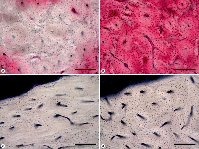
Fig. 1.
Photomicrographs of devitalized bone matrix in the mandible of bisphosphonate-treated dogs. a Matrix necrosis, assessed using en bloc basic fuchsin staining, can be observed in the mandible of a dog treated daily for 1 year with oral alendronate, by the complete absence of fuchsin stain (red) in a localized region. b A region from the mandible of a vehicle-treated animal shows staining of viable tissue. c Focal loss of viable osteocytes, assessed using lactate dehydrogenase histochemistry which labels viable osteocytes blue, can be observed in the alveolar bone of an animal treated for 3 months with intravenous zoledronate. d A region from a vehicle-treated animal shows the majority of osteocytes are viable (stained blue). Scale bars = 200 μm.

