Fig. 5.
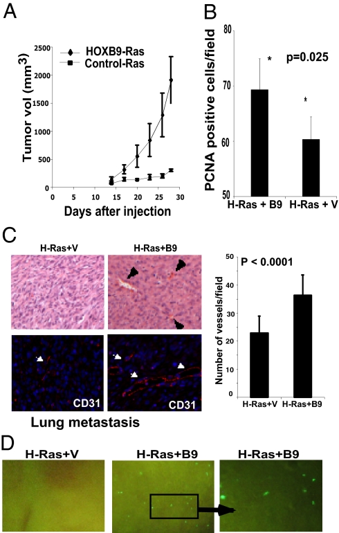
HOXB9 expression promotes tumor growth, angiogenesis, and distal metastasis to the lung. (A) MCF10A cells expressing activated H-Ras alone or activated H-Ras + HOXB9 were inoculated s.c. into mice; the mean change in tumor volume ± SD for each group is shown (n = 8 mice per group). (B) HOXB9-expressing tumors exhibit a higher proliferation index. Tumors from the two groups were stained with antibody to the proliferation marker PCNA. The mean number of PCNA-positive cells ± SD per field is shown (n = 10 fields). (C) HOXB9+activated-Ras tumors exhibit increased vascularization. Vessels are indicted with arrowheads. Quantification of vessels by CD31 staining is shown below. (Original magnification 200×) (D) HOXB9 expression promotes lung metastasis. Lungs of activated H-Ras+HOXB9 tumor-bearing mice (50%) demonstrate micrometastases, whereas none of the animals (0%) bearing activated H-Ras tumors show signs of lung metastasis. The GFP-expressing cell clusters in the lung were visualized under a dissecting microscope. The right panel shows a higher-magnification image of the inset. (Original magnification 40×).

