
Figure 2. Genta stained paraffin sections of a patient with duodenal ulcer (Original magnification ×1,000).
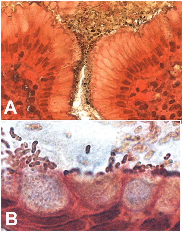
A. Illustration of the presence of dense colonies in the adherent mucus that covers the superficial mucosa of the antral lumen and foveola.
B. Enlargement detail of an antral gland, illustrating the presence of H. pylori colonizing the mucus with characteristic denser colonies at an intercellular junction on the left of the picture; note that H. pylori density overlying each cell is higher near the tallest epithelial cells, when the mucus is being released, and that some H. pylori are adjacent to mucus secretory granules while other bacteria congregate at the apex of the cells.