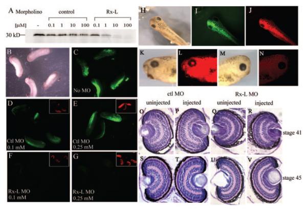
Figure 4.
Injection of the Rx-L anti-sense MO oligonucleotide specifically inhibited translation of Rx-L but did not result in gross changes in eye morphology. (A) The Rx-L antisense MO oligonucleotide (Rx-L MO), but not the control MO oligonucleotide (ctl MO), can inhibit Rx-L translation in vitro. (B–G) The Rx-L MO, but not a control MO, can inhibit expression of a Rx-L-GFP fusion protein containing the Rx-L MO target in Xenopus embryos. (B, C) bright-field (B) and fluorescent (C) views of embryos injected with Rx-L-GFP plasmid. (D, E) GFP fluorescence in embryos injected with Rx-L-GFP plasmid and 0.1 (D) or 0.25 (E) mM ctl MO. (F, G) GFP fluorescence in embryos injected with Rx-L-GFP plasmid and 0.1 (F) or 0.25 (G) mM Rx-L MO. Insets: visualization of lissamine tag of MO (red fluorescence). (H–N) Microinjection of 0.25 mM Rx-L MO did not affect external morphology of the eye. Embryos were unilaterally co-injected with lissamine-conjugated Rx-L MO and GFP RNA at the four-cell stage. (H–J) Dorsal views of injected embryos with bright-field illumination (H), green fluorescence to detect GFP tracer (I) or red fluorescence to detect lissamine-conjugated MO (J). (K, L) lateral view of the injected side of the same embryo viewed with bright-field illumination (K) or red fluorescence (L). (M, N) lateral view of the uninjected side of the same embryo viewed with bright-field illumination (M) or red fluorescence (N). (O–V) Retinas from embryos injected with Rx-L MO appeared histologically similar to those from embryos injected with ctl MO at st 41 and 45 (as indicated).

