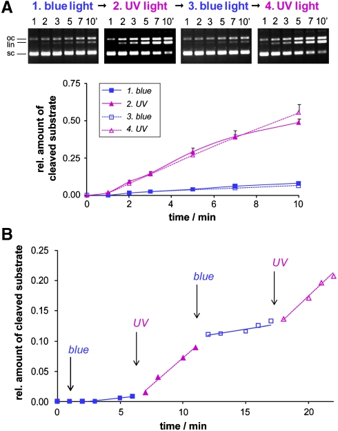
Fig. 3.
(A) Activity changes of sc(C49 C62)2Azo induced by preillumination with blue light → UV light → blue light → UV light as determined by a subsequent DNA cleavage assay (4 nM DNA, 0.5 nM enzyme) under illumination with blue or UV light, respectively. On the Top the gel electrophoretic analysis of samples withdrawn from the incubation mixture at defined time interval is shown, on the Bottom the corresponding activity vs. time of illumination profile. According to the quantitative analysis, sc(C49 C62)2Azo is seven times more active with the azobenzene moiety in the cis- than in the trans-configuration. (B) Reversible photoswitching of the DNA cleavage activity of sc(C49 C62)2Azo observed in situ. After preilluminating the enzyme with blue light, DNA cleavage (4 nM DNA, 0.24 nM enzyme) was followed in situ with variation of the illumination during cleavage (blue light 0–6 min, UV light 6–11 min, blue light 11–17 min, UV light 17–22 min). sc, oc, and li denote the supercoiled, open circular, and linear forms, respectively, of the plasmid DNA substrate.

