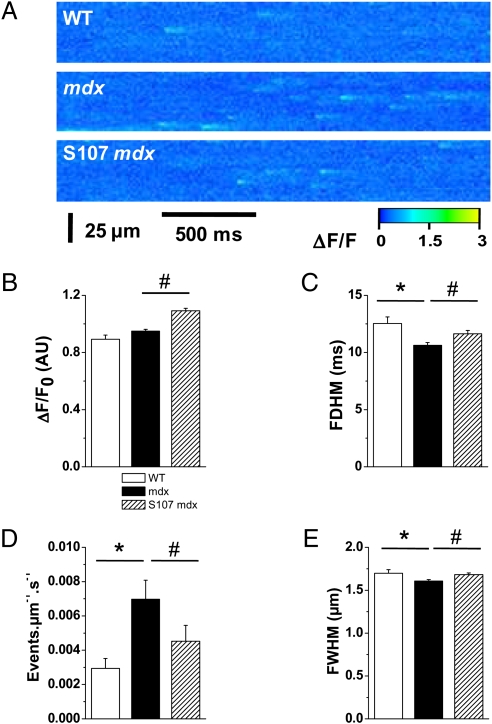
Fig. 2.
SR Ca2+ leak assessed by Ca2+ sparks analysis in mdx mice. Spontaneous SR Ca2+ release events were recorded in fluo-4 AM–loaded intact cardiomyocytes by laser scanning confocal microscopy, as described in SI Materials and Methods. (A) Representative ΔF/F line scan images (1.54 ms/line) were recorded in WT (Top) and mdx mice without (Middle) and with (Bottom) S107 treatment. Diastolic SR Ca2+ leak is estimated by the average sparks frequency (D). Average spatiotemporal properties of Ca2+ sparks, such as amplitude (B), full duration at half maximum (C), and spatial spread (full width at half maximum) (E). Data are expressed as mean ± SEM. *P < 0.05 WT vs. mdx; #P < 0.05 mdx vs. S107-mdx. n = 195 sparks in 20 cells in WT, 1,272 sparks in 58 cells in mdx, and 889 sparks in 60 cells in S107-treated mdx mice.

