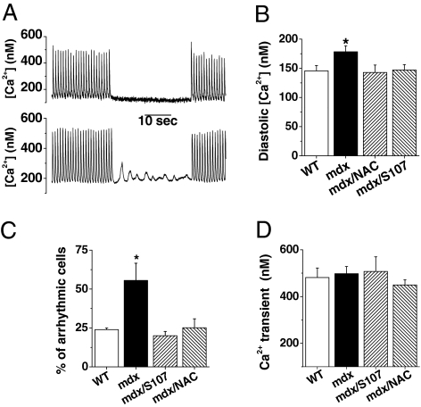
Fig. 3.
Elevated diastolic Ca2+ concentration in mdx mice. Isolated cardiomyocytes, loaded with indo-1 AM as described in SI Materials and Methods, were paced at 1 Hz. After 1 min, cells were maintained quiescent for 30 s before a second train of stimulation. (A) Global Ca2+ was recorded simultaneously, as illustrated by two typical recordings in WT and in mdx cardiomyocytes. (B) During the stimulation, diastolic Ca2+ was elevated in mdx cardiomyocyte. This was prevented by NAC or S107 treatment. (C) During the 30-s rest period, ∼50% of mdx cardiomyocytes exhibited Ca2+ waves that were not observed in WT or in mdx after NAC or S107 treatment. (D) The peak amplitude of the Ca2+ transients did not differ significantly in all conditions. Data are expressed as mean ± SEM. WT, n = 24; mdx, n = 29; mdx-S107, n = 15; mdx-NAC, n = 28. *P < 0.05.

