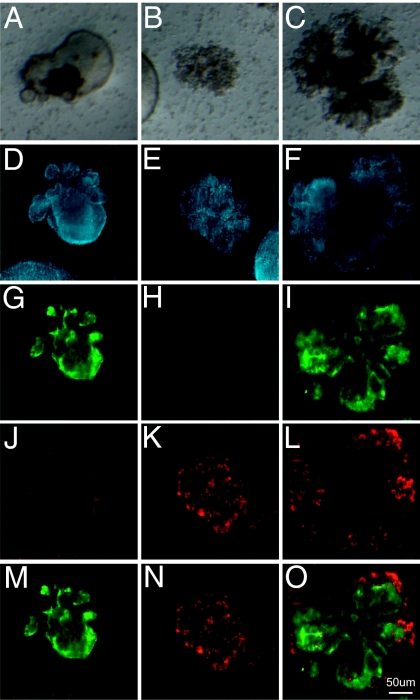
Fig. 4.
Generation of distinct epithelial colony subtypes. Bright-field images of lobular cystic airway-like colonies (A), dense saccular alveolar-like colonies (B), and colonies with mixed morphologies (C). Fluorescent confocal images of DAPI (blue) (D–F), MUC5AC (green) (G–I), proSP-C (red) (J–L), and overlay of MUC5AC and proSP-C staining of representative colonies (M–O).

