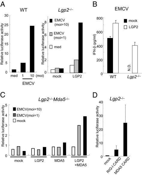
Fig. 3.
LGP2 acts in the upstream of RIG-I and MDA5. (A) WT and Lgp2 −/− MEFs were transiently transfected with the IFN-β promoter construct together with expression plasmids encoding LGP2. The cells were infected with EMCV for 8 h and then lysed. The cell lysates were analyzed by a luciferase assay. (B) WT and Lgp2 −/− MEFs were infected with a retrovirus expressing Lgp2. At 2 days after infection, the cells were exposed to EMCV for 24 h. The IFN-β concentrations in the culture supernatants were measured by ELISA. N.D., not detected. (C) Lgp2 −/− Mda5 −/− MEFs were transiently transfected with the IFN-β promoter reporter construct together with the indicated expression plasmids. After 24 h, the cells were infected with EMCV for 8 h and then lysed. The cell lysates were analyzed by a luciferase assay. (D) Lgp2 −/− MEFs were transiently transfected with the IFN-β promoter construct together with expression plasmids encoding the CARDs of RIG-I or MDA5 and then lysed at 48 h after transfection. The cell lysates were analyzed by a luciferase assay.

