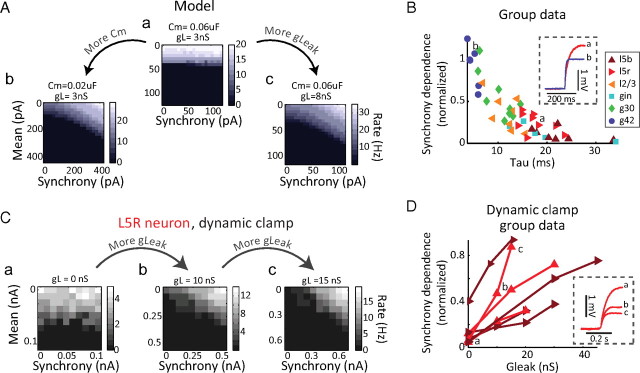
Figure 4.
The relative synchrony dependence of neurons depends on passive membrane-filtering properties. A, The response of a single-compartment Hodgkin–Huxley-style model neuron to the stimulation protocol is relatively synchrony independent. a, Decreasing its time constant either by decreasing the capacitance (a→b) or increasing the leak conductance (a→c) increases synchrony dependence. B, Across n = 47 cells, time constant correlates with normalized synchrony dependence (Spearman's r = −0.84; p = 4.11 e-013). An example voltage trace from a layer 5 regular-spiking neuron with a short time constant (a) and an example G42 fast-spiking with a long time constant (b) are illustrated and corresponding points marked. Neuron types are color- and marker-coded as in Figure 3. C, Changing membrane-filtering properties changes synchrony dependence. The response of an example L5 regular-spiking cell is weakly dependent on input synchrony; reducing its time constant by the addition of leak conductance (a→b→c) increases its relative synchrony dependence. D, Across n = 7 L5R (red) and L5B (dark red) cells, increasing the leak conductance reliably increased the synchrony-to-mean ratio of the neuron (p < 0.05). Trial blocks shown in inset are marked. Inset, Example voltage traces from Ca, Cb, and Cc.

