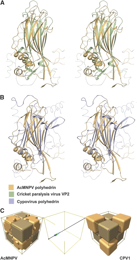
Figure 2.
Structural superpositions. Stereo views showing the alignment of (A) AcMNPV polyhedrin (orange) with cricket paralysis virus VP2 (green) (Tate et al, 1999) and (B) AcMNPV polyhedrin (orange) with CPV1 polyhedrin (purple). The program SHP (Stuart et al, 1979) was used to perform the superpositions. (C) Schematic representation of the packing of both AcMNPV and CPV1 polyhedrin trimers (shown as blocks) within their unit cells. The trimeric units are positioned rather differently on the three-fold body diagonal—this is represented in the central panel in which the centres of the trimers of differing polarity are reflected into the (0,0,0) to (½, ½, ½) portion. The AcMNPV trimer centres (shown in shades of green) lie close to (¼, ¼, ¼), whereas the CPV1 trimers (centres shown in shades of blue) are offset by about 6 Å. This accounts for the differences seen in the outer panels.

