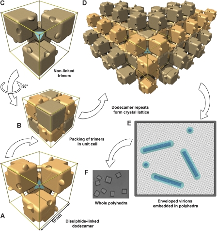
Figure 4.
Schematic representations of polyhedra organization. Polyhedrin trimers are depicted as simplified cubic blocks, with the C-terminal hooks and pockets present. To clarify interpretation, the edges of the unit cell are shown in gold and a cyan tetrahedron symbolizes the cell centre. Within a unit cell, disulphide-linked trimers with one polarity are coloured light beech (A) and those with the opposite polarity are coloured light brown (C). (B) All eight trimers in the unit cell. The disulphide bond connecting adjoining trimers is shown as a dowel. (D) The crystal lattice is built up from repeats of the dodecameric unit. (E) Sketch of a cross-section through a polyhedron. The lattice spacing of the unit cells is illustrated as a dot pattern into which are embedded nucleocapsids (dark blue) surrounded by an envelope (cyan). (F) Light microscopy image of G25D mutant AcMNPV polyhedra.

