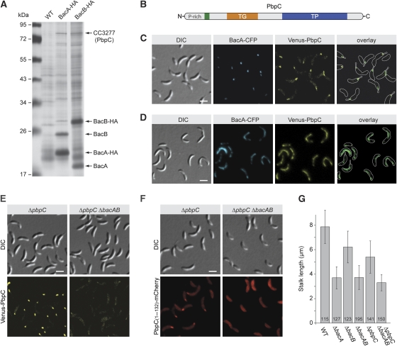
Figure 5.
Interaction of BacA and BacB with the penicillin-binding protein PbpC. (A) Identification of proteins interacting with BacA and BacB. Lysates prepared from formaldehyde-treated cells of strains CB15N (wild type), KL7 (bacA-HA), and KL8 (bacB-HA) were subjected to co-immunoprecipitation analysis using anti-HA-affinity beads. Precipitated proteins were resolved in an 11% SDS–polyacrylamide gel and detected by silver staining. Bands specific for the samples from strains KL7 or KL8 were analysed by mass spectrometry. Owing to the limited amount of starting material, only the five most abundant proteins could be identified. (B) Schematic representation of PbpC. The transmembrane helix (residues 86–108) is indicated in green, the transglycosylase domain (TG; residues 133–300) in orange, and the transpeptidase domain (TP; residues 401–668) in blue. (C) Colocalization of BacA and PbpC. Cells of strain JK271 (bacA-ecfp xylX::Pxyl-venus-pbpC) were grown in PYE-rich medium, induced for 1 h with 0.03% xylose, and visualized by DIC and fluorescence microscopy (bar: 2 μm). (D) Detection of an interaction between BacA and PbpC in vivo. Cells of strain MT279 (xylX::Pxyl-venus-pbpC) carrying overexpression plasmid pJK53 (Pxyl-bacA-ecfp) were grown in PYE-rich medium, induced for 4 h with 0.3% xylose, and visualized by DIC and fluorescence microscopy (bar: 2 μm). Note: For strain MT279 lacking an overexpression plasmid, only polar signals were observed when grown under the same conditions (data not shown). (E) Loss of polar PbpC localization on deletion of bacA and bacB. Strains JK308 (ΔpbpC xylX::Pxyl-venus-pbpC) and JK310 (ΔbacAB ΔpbpC xylX::Pxyl-venus-pbpC) were grown in PYE-rich medium, induced for 1 h with 0.03% xylose, and visualized by DIC and fluorescence microscopy (bar: 2 μm). (F) Determination of the PbpC region responsible for interaction with BacA and BacB. Strains JK308 (ΔpbpC xylX::Pxyl-pbpC[AA 1−132]-mCherry) and JK289 (ΔbacAB ΔpbpC xylX::Pxyl-pbpC[AA 1−132]-mCherry) were grown in PYE-rich medium, induced for 2 h with 0.03% xylose, and visualized by DIC and fluorescence microscopy (bar: 2 μm). (G) Reduced stalk length of strains lacking bactofilin homologues and/or PbpC. Strains CB15N (wild type (WT)), MT257 (ΔbacA), MT259 (ΔbacB), JK5 (ΔbacAB), MT304 (ΔpbpC), and JK281 (ΔbacAB ΔpbpC) were grown in PYE-rich medium, diluted 1:20 in minimal medium lacking phosphate (M2G−P), and cultivated for another 24 h. The graph gives the average stalk length (±s.d.) reached under these conditions and the number of cells analysed.

