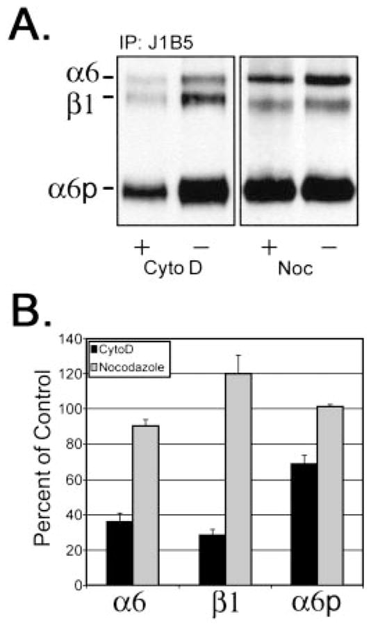
Fig. 3.

Disruption of the actin cytoskeleton significantly reduced cell surface expression of α6, α6p, and β1 integrins. Surface changes of α6, β1, and α6p were determined by surface of DU145H cells with biotin before treatment with either 10 μM cytochalasin D or 8 μM nocodazole for 18 h. Labeled cells were lysed, and 200 μg of total protein were used for immunoprecipitations with anti-α6 integrin antibody, J1B5. Samples were separated on a 7.5% polyacrylamide gel under nonreducing conditions. Proteins were transferred to PVDF membrane followed by incubation with HRP conjugated to streptavidin (A). Resulting α6, β1, and α6p integrin protein bands were quantified and graphed (B).
