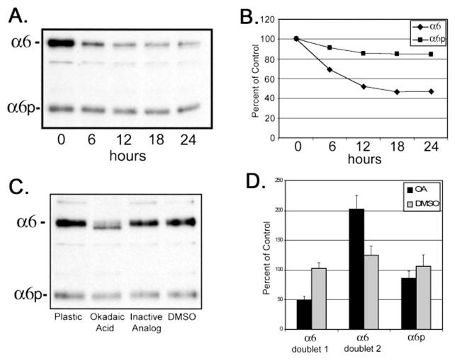
Fig. 5.

Calyculin A and okadaic acid treatment of DU145H cells decreased α6 integrin protein levels but not α6p. Human prostate carcinoma DU145H cells were treated with 15 nM calyculin A over a 24-h period of time. Identical amounts of whole cell lysate (10) were loaded and electrophoresed on a 7.5% polyacrylamide gel under nonreducing conditions. Proteins were transferred to PVDF membrane followed by Western analysis for α6 integrin (A). Protein bands in A were scanned and quantified using Scion Image Analysis software and graphed in Excel (B). Data shown were representative of three independent experiments. DU145H cells were treated with 50 μM okadaic acid, the inactive analogue 1-nor-okadaone, or vehicle (DMSO) for 18 h. Whole cell lysates were examined for α6 integrin protein expression as above (C). Resulting α6 and α6p bands from three independent experiments were quantified and graphed (D).
