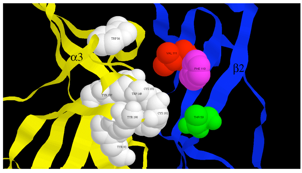
Fig. 8.
α3β2 nAChR binding site. The ACh binding site at an α3/β2 interface is shown within a model of the α3β2 extracellular domain. Proposed α3 subunit agonist binding residues (33) shown in white space filling. Residues affecting α-conotoxin BuIA binding kinetic differences between β2 and β4 subunits shown in color space filling.

