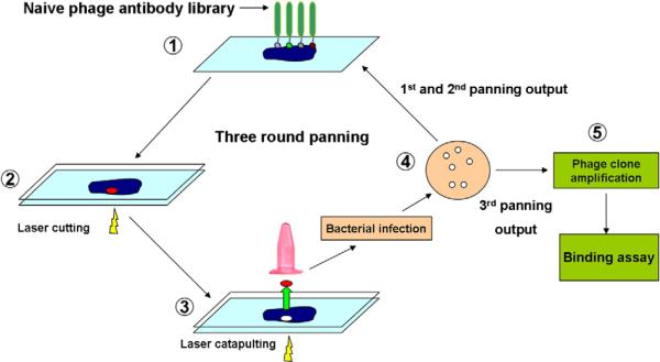
Fig. 1.
Schematic presentation of phage panning on tissues sections with LCM. Naive phage library was directly incubated with tissue sections mounted on PALM membrane slides (1). After rinsing steps, a blank PALM membrane covered slides to keep tissue moist (2). Tissue was cut (2) and captured (3) by laser beams. The collected tissue pieces were directly used to infect host stain, and infected cells were selected on TYE-ampicillin plate (4). Panning outputs were pooled and amplified for the next round of panning. Three rounds of panning were performed and individual colonies were randomly selected from the 3rd panning output for binding assay (5).

