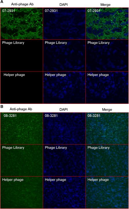
Fig. 3.
Immunofluorescence analysis of phage clone 07-2931 (A) and clone 08-3281 (B) binding to human breast cancer tissue. Breast cancer frozen sections derived from the same clinic patient for panning were incubated with phage clone while same quantity phage library and no-insert helper phage were usedas control. The phage was visualized by fluorescence using mouse-anti-M13 phage antibody followed by Alex-488-conjugated goat anti-mouse antibody. DAPI was used for nuclear counterstaining and tissue structure. Immunofluorence (IF) stain with phage clone (first row of A and B) show selective signals (green) located on tumor stroma (A) and tumor cells (B). As controls, Naive scFv phage library stain (second row) and M13 helper phage (third row) show no signals on tumor tissue.

