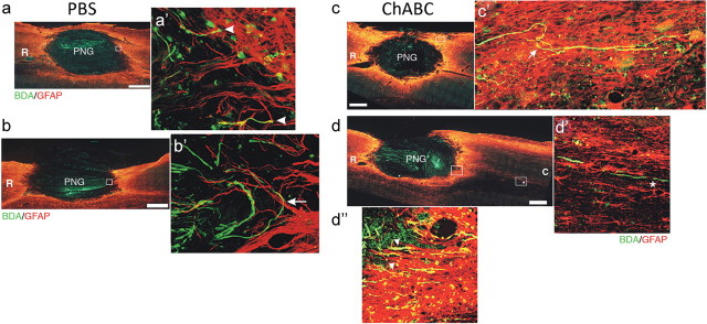
Figure 6.
Chondroitinase ABC treatment allows long-injured axons to extend beyond a peripheral nerve graft spanning a cavity. Horizontal sections of hemicontused C5 spinal cord containing a PNG are depicted. Chronically injured axons that grew into the PNG were labeled with BDA (green). GFAP+ astrocytes (red) demark the boundary of host spinal cord tissue and the graft. In PBS-treated tissue (n = 9), BDA+ axons were able to grow up to the distal interface but failed to extend beyond it (a, b). Rather, they displayed hallmarks of regenerative failure, including bulbous, dystrophic endings (a′, arrowheads), and turning at the inhibitory boundary (b′, arrow). Conversely, ChABC treatment allowed many chronically injured axons to cross the interface (d″, arrowheads; n = 12) into astrocyte-rich territory. Some axons took a cursive course through host tissue (c′, arrow), indicating that they regenerated. BDA+ fibers were found several millimeters away from the distal interface (d′, asterisk). Significantly more BDA+ fibers were found in tissue 100 μm caudal to the distal interface after ChABC treatment (37.2 ± 5.7 axons per 10 sections) than PBS treatment (5.8 ± 1.5 axons per 10 sections; p = 0.0001). Insets a′, b′, and c′ are higher-magnification images of the boxed regions in a, b, and c, respectively. d′ is a high-magnification image of the rightmost boxed region in caudal tissue (C) in d. d″ is a high-magnification image of the boxed region at the interface in d. R, Rostral. Scale bar, 500 μm.

