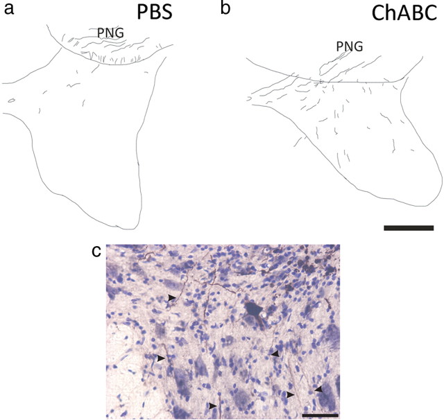
Figure 7.
Treatment with chondroitinase ABC promotes chronically injured axons to regenerate out of a peripheral nerve graft bridge. Transverse sections of spinal cord containing a PN apposed to a C7DQ injury were reacted with DAB to visualize BDA-labeled chronically injured axons that grew into the PNG. Tracings of representative montages are represented in a and b. Few BDA+ axons were seen in tissue 500 μm ventral to the graft (a) after PBS treatment (1.4 ± 0.36 axons per section; n = 10). However, after administration of ChABC (n = 6), many more labeled axons regenerated out of the graft (b; 6.5 ± 1.0 axons per section; p = 0.00005) and could be found in ventral, ipsilateral tissue. A high-magnification image of BDA+ axons (highlighted by arrowheads) extending into ventral, ipsilateral gray matter of a representative, ChABC-treated section is shown in c. Scale bars: (in b) a, b, 250 μm; c, 50 μm.

