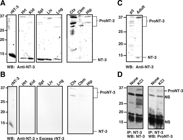
Figure 2.
Neuronal release of endogenous proNT-3. A, Tissue-specific expression of proNT-3 and mature NT-3. Detergent lysates were prepared from the indicated tissues. Except for heart and kidney in which 100 μg of the respective lysates were analyzed, equal amounts of visceral tissue lysates (50 μg) and 150 μg of CNS tissue lysates were Western blotted with an anti-NT-3 antiserum (sc-547). This was done because the range of proNT-3/NT-3 expression varies greatly between these tissues (Kaisho et al., 1994; Katoh-Semba et al., 1996); the resulting enhanced chemiluminescence signals would have exceeded the linearity of the film without the loading adjustment. Note that various non-neural and CNS tissues express mature NT-3 and proNT-3 at varying proportion and abundance. The numbers on the left indicate positions of the molecular weight markers. The brackets on the right indicate multiple proNT-3 species whose identities were confirmed by reprobing the blot with anti-proNT-3 antiserum and an independently generated anti-NT-3 antibody (data not shown). Ctx, Cortex; Cbm, cerebellum; Hip, hippocampus; Spl, spleen; Hrt, heart; Lng, lung; Kid, kidney; Liv, liver. B, To demonstrate specificity of the proNT-3 and mature NT-3 species, anti-NT-3 antibody (sc-547) was preincubated with excess recombinant NT-3 before addition to a replica tissue lysates blot but was otherwise processed in identical manner as in Figure 2A. C, Age-dependent expression of proNT-3 and mature NT-3. Cerebellar lysates from early postnatal (P3) and adult female rat were Western blotted with anti-NT-3 (sc-547). Positions of proNT-3 and mature NT-3 are indicated on the right. D, P6 cerebellar granule neurons in culture were treated with either 5 mm KCl (None) or with 25 mm KCl for 24 h in the presence of a goat anti-NT-3 antibody (sc-13380) as described in Materials and Methods. Anti-NT-3 immunoprecipitates were Western blotted with either rabbit anti-NT-3 antiserum (sc-547; left panel) or with anti-proNT-3 specific antiserum (right panel) to verify the identify of proNT-3. The position of proNT-3 is indicated on the left as are positions of nonspecific bands (NS).

