
Figure 7.
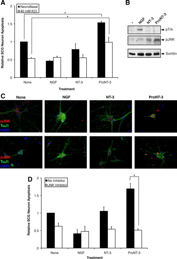
A role for JNK in proNT-3-induced neuronal apoptosis. A, Replica cultures of rat SCG neurons (DIV 7) were washed free of NGF and were treated or not with 10 ng/ml NGF or equal molar NT-3 or proNT-3 (i.e., 2 ng/ml NT-3 or 4 ng/ml proNT-3) in the presence or absence of 40 mm KCl as indicated. Neuronal apoptosis was assessed for each culture condition 48 h later. The data, normalized to the number of apoptotic neurons on NGF deprivation, represent values from three independently conducted experiments. The vertical error bars indicate SEM. Note that membrane depolarization prevented NGF-deprived neuron death (Franklin et al., 1995) but not did not affect proNT-3-induced apoptosis. B, Replica cultures of rat SCG neurons (DIV 7) were washed free of NGF and were then treated with 12.5 mm KCl as described for analysis of JNK activation (Linggi et al., 2005). SCG neurons were treated or not with 50 ng/ml NGF, 25 ng/ml mature NT-3, or 25 ng/ml proNT-3 as indicated for 4 h. Cellular lysates (100 μg each) were Western blotted with the indicated antisera to assess proNT-3-induced JNK activation relative to TrkA-specific signaling. Equality of sample loading was verified by reprobing the blot with an anti-sortilin antiserum (BD Biosciences). C, Replica cultures of P1 SCG neurons (DIV 7) were washed free of NGF and maintained in 12.5 mm KCl either in presence or absence of NGF, NT-3, or proNT-3 as indicated. Forty-eight hours later, cells were fixed and stained with TuJ1 (Covance), pJNK (Cell Signaling), and DAPI as indicated. Only proNT-3-treated, apoptotic neurons exhibited pJNK immunoreactivity (top panels). Alternatively, cultures were stained with TuJ1 and pJun as corroborative evidence of JNK activation (bottom panels). D, Replica cultures of rat SCG neurons (DIV 7) were washed free of NGF and were treated or not with 10 ng/ml NGF or equal molar of NT-3 or proNT-3 (i.e., 2 ng/ml NT-3 or 4 ng/ml proNT-3) in the presence or absence of 15 μm SP600125 as indicated. Neuronal apoptosis under standard (non-depolarizing) culture condition were assessed 48 h after treatment. The data, normalized to the number of apoptotic neurons on NGF deprivation, represent values from three independently conducted experiments, and the vertical error bars indicate SEM. For A and D, asterisks denote statistical significance between the paired samples.