
Figure 8.
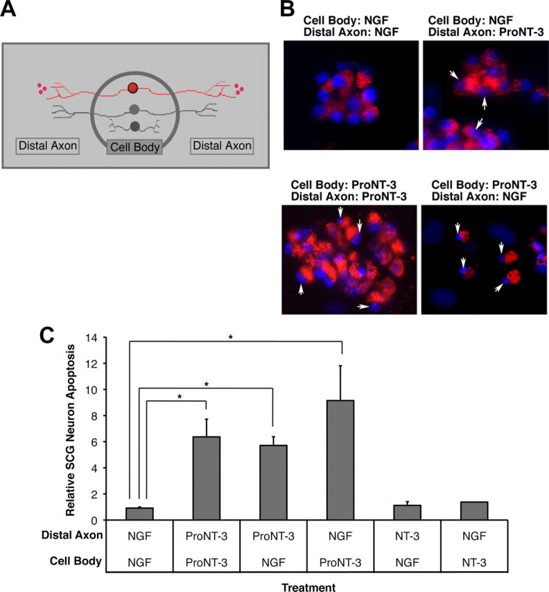
Retrograde apoptotic signaling. A, Schematic representation of compartmentalized culture system. Only neurons (red) that were prelabeled with fluorescent microspheres were evaluated in subsequent analysis. B, Fluorescent photomicrograph of SCG neurons under the indicated culture conditions. Only viable neurons with active retrograde transport take up fluorescent microspheres from the distal axon compartment. The arrows indicate fragmented nuclei of dying neurons. C, Assessment of neuronal apoptosis under each culture conditions. Replica compartmentalized cultures were treated with 1 ng/ml NGF or equal molar of NT-3 or proNT-3 (i.e., 5 ng/ml NT-3 or 10 ng/ml proNT-3) in the center cell body compartment as indicated, whereas 10 ng/ml NGF or equal molar of NT-3 or proNT-3 (i.e., 5 ng/ml NT-3 or 10 ng/ml proNT-3) was applied to the distal axon compartment. Neuronal apoptosis was evaluated 48 h later. The data, normalized to the number of apoptotic neurons maintained with NGF in both compartments, were pooled from five independently conducted experiments. The vertical error bars indicate SEM. Asterisks denote statistical significance between the paired samples.