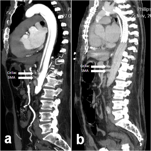
Figure 2.

(a) Pre-operative sagittal view of the thoracic aorta showing contrast within the true and false lumina. Note near total occlusion of the celiac and superior mesenteric arteries (SMA) denoted by white arrows. (b) Post operative sagittal image of the same aortic segment with stent graft in-situ demonstrating increased flow within the celiac and superior mesenteric arteries (SMA) denoted by white arrows.
