
FIGURE 2. IL-1 activates NEMO-dependent classical NF-κB in IKKβ−/− MEFs.
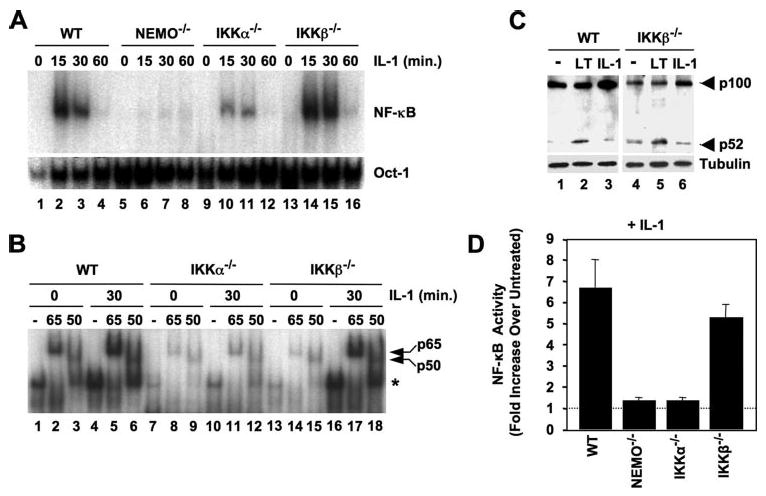
A, WT, NEMO-deficient, IKKα−/−, and IKKβ−/− MEFs were either untreated or incubated with IL-1 for the times indicated, and then nuclear extracts were prepared and used for EMSA. Assays were performed using either a consensus NF-κB binding site probe (upper panel) or an Oct1 probe as a loading control (lower panel). B, the MEFs shown (top) were either untreated or incubated with IL-1 for 30 min, and then nuclear lysates were prepared. For supershift analysis, samples were incubated prior to the EMSA reaction either in the absence of antibodies (−) or with anti-p65 or -p50 as shown. The positions of the shifted NF-κB complex (*) and supershifted p65- and p50-containing complexes are indicated (right). C, WT and IKKβ−/− MEFs were either untreated (−) or stimulated with anti-LTβR (LT) or IL-1 (10 ng/ml) for 8 h, and then lysates were immunoblotted and probed with either anti-p100/p52 (upper panel) or anti-tubulin (lower panel). D, MEFs were transiently transfected with the NF-κB-dependent reporter pBIIx-firefly luciferase together with β-actin Renilla luciferase. Twenty-four hours later, the cells were either untreated or incubated for a further 5 h with IL-1 (10 ng/ml), and then NF-κB activity was determined by dual luciferase assay. The data are expressed for each MEF line as fold values relative to the basal activity in untreated cells that was normalized between the cell lines (dotted line).