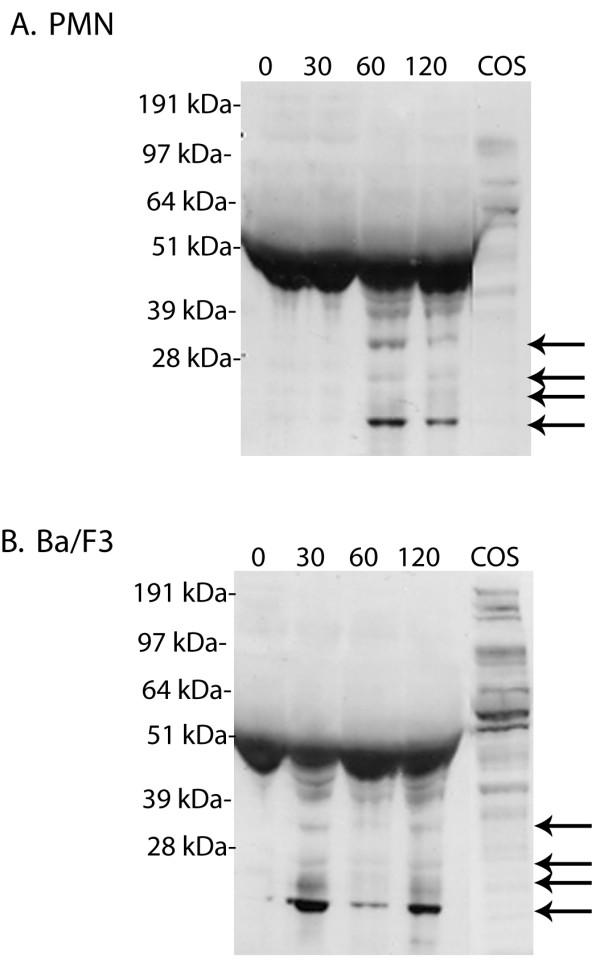
Figure 3.

Detection of G-CSFR cleavage fragments in conditioned media from NE-treated cells. (A) PMN and (B) Ba/F3 cells transfected with the full-length G-CSFR cDNA were treated (1 × 10⁷cells) with 150 μg/ml NE for the indicated times. Reactions were stopped by the addition of 10% FBS and 1 mM PMSF, the samples centrifuged, and the supernatants containing the conditioned media collected and immunoblotted with an antibody recognizing the FNIII domains in the extracellular region of the G-CSFR. Arrows indicate the extracellular G-CSFR fragments generated by NE. A representative blot from three independent experiments (n = 3) is shown.
