
FIGURE 6.
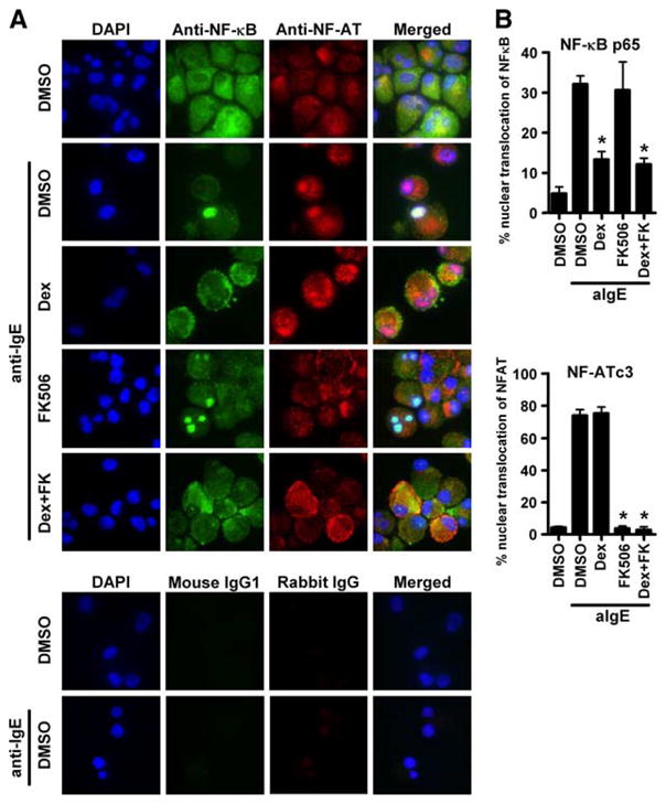
Effect of FK506 and DEX on the translocation of NF-κB and NF-AT by anti-IgE treatment in human mast cells. IgE-sensitized human mast cells were preincubated with either 1 μM DEX, 100 nM FK506, or 0.01% DMSO for 1 h and then stimulated with 1.5 μg/ml anti-IgE Ab for 30 min. A, Immunofluorescence images were showing the distribution of NF-κB and NF-AT. Mast cells were treated with mouse anti-NF-κB p65 and rabbit anti-NF-ATc3 for localization of endogenous NF-κB (green fluorescence) and NF-AT (red fluorescence). Nuclei were counterstained with 4′,6-diamidino-2-phenylindole (DAPI) (blue fluorescence). The images are representative of four independent preparations. B, Summary of the percentage of mast cells in which NF-κB p65 (upper panel) and NF-ATc3 (lower panel) has localized within the nuclei. The results are shown as the means ± SEM of four independent experiments.*, p < 0.05.