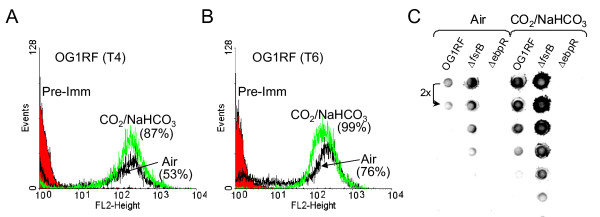
Figure 3.
Detection of EbpC produced by OG1RF, ΔfsrB, and ΔebpR. A. Flow cytometry analysis of OG1RF grown in air (black) or in the presence of 5% CO2/0.1 M NaHCO3 (green) labeled with an anti-EbpC rabbit polyclonal immune serum and detected with phycoerythrin. The cells were collected at "T4", which corresponds to the entry into stationary growth phase (4 hrs after starting the culture). The percentages between brackets indicate the percentage of positive cells (WinMDI 2.9, marker set for 500-1024). In red is represented OG1RF grown in air incubated with a pre-immune serum and detected with Phycoerythrin as negative control. B. Flow cytometry analysis was done in the same conditions as above with samples collected at "T6" which corresponds to early stationary growth phase. C. An equal amount (by BCA protein assay) of mutanolysin extract preparation was 2-fold serial diluted and spotted onto a nitrocellulose membrane. Pilus presence was detected with an anti-EbpC rabbit polyclonal immune serum.

