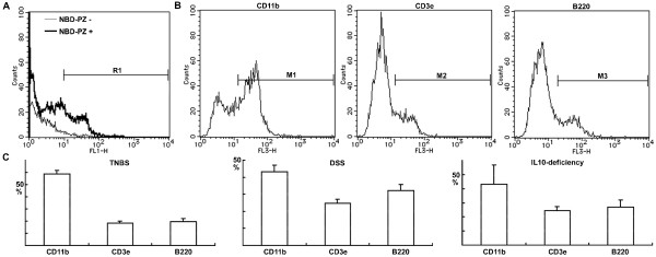
Figure 5.
Characterization of NBD-PZ-stained mononuclear cells isolated from the mucosa of colitis. (A) Mononuclear cells were isolated from the mucosa of TNBS-induced colitis and incubated with 0.25 μg/ml NBD-PZ at 37°C for three minutes for flow cytometry. A total of 10,000 cells were analyzed. The cells in the R1 region were defined as NBD-PZ-positive. (B) Mononuclear cells were isolated from the mucosa of TNBS-induced colitis, incubated with 0.25 μg/ml NBD-PZ at 37°C for three minutes, and reacted with anti-CD11b, CD3e or B220 antibody on ice for 30 minutes. A total of 10,000 NBD-PZ-stained cells were analyzed by flow cytometry and the cells in the M1, M2 or M3 region were defined as CD11b-, CD3e- or B220-positive, respectively. (C) Mononuclear cells were isolated from the mucosa of colitis induced by TNBS, DSS or IL-10 deficiency to characterize the NBD-PZ-stained cells under flow cytometry. Data represent the percentages of CD11b-, CD3e- and B220-positive cells within the NBD-PZ-stained cell pool, respectively (means ± standard deviations, n = 3).

