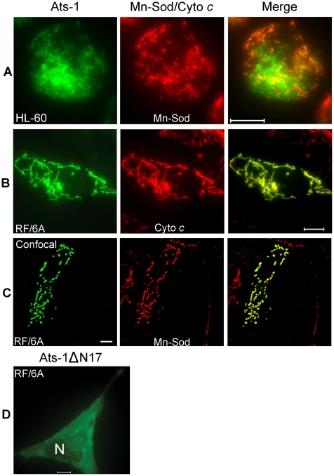
Figure 4. Mitochondrial targeting of Ats-1, and essential role of N-terminal sequence in targeting.
A-C. Double immunofluorescence labeling of A. phagocytophilum–infected HL-60 cells (A), or pAts-1-transfected RF/6A cells (B and C) using rabbit anti-Ats-1 (Ats-1; Alexa Fluor 488, green), and monoclonal anti-Mn-Sod (Mn-Sod; Alexa Fluor 555, red) (A and C), monoclonal anti-cytochrome c (Cyto c; Alexa Fluor 555, red) (B). Scale bar: 10 µm. D. Immunofluorescence labeling of RF/6A cells transfected with pAts-1ΔN17 was performed using anti–Ats-1 (Alexa Fluor 488, green). N, nucleus. Note diffuse distribution of Ats-1ΔN17 in the cytoplasm of RF/6A cell. Scale bar: 10 µm.

