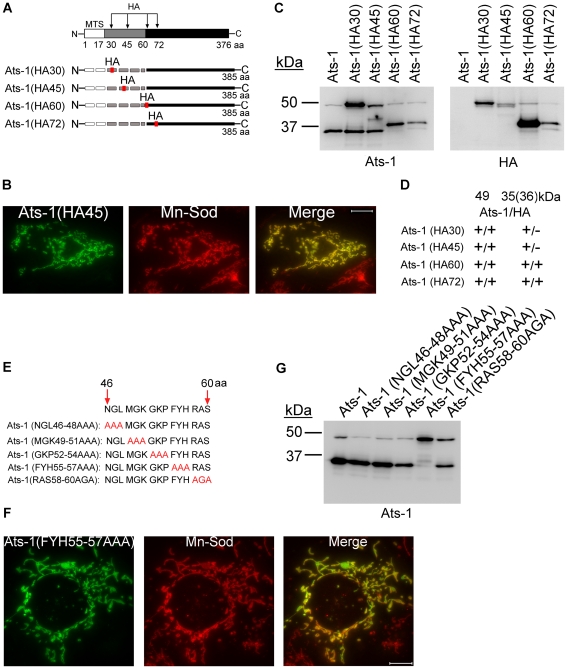
Figure 6. Ats-1 presequence is cleaved at a specific site.
A. Schematic diagram indicating HA tag insertion and predicted cleavage sites in Ats-1 mutants. The HA tag was inserted between residues 30 and 31, 45 and 46, 60 and 61, or 72 and 73 of Ats-1. MTS, predicted mitochondrial targeting sequence, indicated by white bar. Black bar indicates cleaved Ats-1 fragments detected by Western blot analysis; Gray bar indicates the sequence between MTS and cleavage site. Dashed lines indicate undetectable (degraded) N-terminal cleaved fragment. B. Immunofluorescence labeling of RF/6A cells transfected with pAts-1 (HA45) using rabbit anti-Ats-1 [Ats-1(HA45); Alexa Fluor 488, green] and mouse monoclonal anti-Mn-Sod (Mn-Sod; Alexa Fluor 555, red). Note mitochondrial localization of Ats-1 (yellow). Scale bar: 10 µm. C. Western blot analysis using anti-Ats-1 or monoclonal anti-HA antibody was performed to examine Ats-1 cleavage in RF/6A cells transfected with recombinant plasmids encoding wild type Ats-1 or Ats-1 mutants with HA insertion at different locations. Molecular mass markers are indicated at left. D. Summarized result for Figure 6C. The molecular size for four Ats-1 mutants in Western blot analysis using anti-Ats-1 (Ats-1) or anti-HA (HA) antibody for Ats-1 (49 kDa) and cleaved Ats-1 (35, or 36 kDa). + indicates immunoreactivity; - indicates no immunoreactivity. Note the molecular mass of full-length Ats-1 or cleaved Ats-1 which has HA insertion increases 1 kDa. E. Diagram showing sequential substitution of amino acid triplets within residues 46–60 of full-length Ats-1. The wild type Ats-1 sequence at residues 46–60 was shown at the top and the mutant sequences in this region are shown below. Substituted residues were indicated in red. F. Immunofluorescence labeling of RF/6A cells transfected with pAts-1 (FYH55-57AAA) using rabbit anti-Ats-1 [Ats-1(FYH55-57AAA); Alexa Fluor 488, green] and mouse monoclonal anti–Mn-Sod (Mn-Sod; Alexa Fluor 555, red). Note mitochondrial localization of Ats-1 (yellow). Scale bar: 10 µm. G. Western blot analysis using anti–Ats-1 was performed to examine Ats-1 cleavage in RF/6A cells transfected with recombinant plasmids encoding wild type Ats-1 or the indicated Ats-1 triplet substitution mutant. Cleavage was inhibited only in cells expressing the Ats-1 (FYH55-57AAA) mutant. Molecular mass markers are indicated at left.

