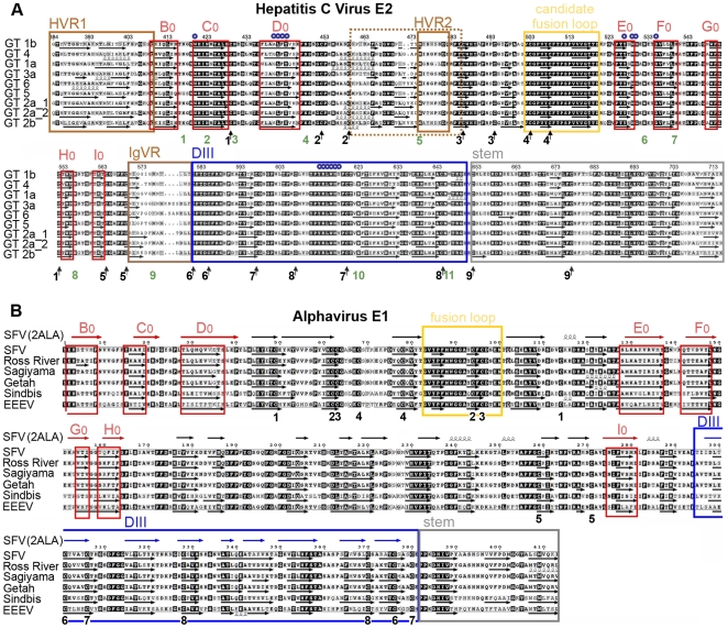
Figure 3. Amino acid sequence alignments and secondary structure predictions.
The secondary structure of HCV E2e and alphavirus sE1 was predicted using the program DSC [48], which was selected because the predictions matched more closely the crystallographically determined secondary structure elements of class II proteins. Arrows or spirals under each sequence indicate predicted β-strands or α-helices, respectively. A) The main elements of the tertiary structure model of HCV E2 are framed: assigned strands in DI (red, labeled) and DIII (blue), putative fusion loop (yellow), the stem (grey) and regions that can be deleted without affecting the protein conformation (brown). The 18 cysteines, which form the 9 disulfides, are marked with arrows and numbered according to the disulfide bond (Table 2) under the sequences. N-linked glycosylation sites are numbered in green. Residues known to interact with CD81 are marked with small blue circles. The numbering corresponds to the HCV H77 polyprotein. B) Comparison with a class II fusion protein of known structure. The experimentally determined secondary structure of SFV E1 taken from the crystal structure (PDB 2ALA, [49]), shown with symbols (arrows or spirals) above the sequence alignment, was compared with secondary structure predictions for the E1 ectodomain of selected alphaviruses. Experimentally determined β-strands in DI, DII and DIII are colored red, black and blue, respectively. The red frames indicate the consensus predicted strands in DI, for easier comparison with panel A. Similarly the fusion loop (yellow), the region corresponding to the crystallographically identified DIII (blue), and the stem (grey) are framed. The 16 cysteines forming the 8 disulfides are numbered in black according to the disulfide bond under the sequences. The numbering starts with the first amino acid of the E1 glycoprotein.

