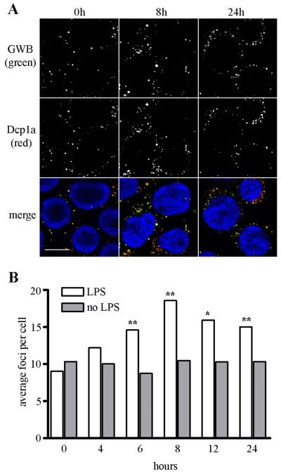
Figure 1. LPS induces the assembly of GWB.
(A) THP-1 cells treated with 1 μg/ml LPS showed a time-dependent increase in the number and size of GWB. After LPS treatment, cells were fixed and costained with human anti-GWB serum (green) and rabbit anti-Dcp1a (red). Nuclei were counterstained by 4,6-diamidino-2-phenylindole (DAPI, blue). Images were acquired at 400x original magnification. Bar, 10 μm. (B) Bar graph showing representative data from the CellProfiler image analysis software used to quantitate the average number of foci per cell in untreated cells or LPS-treated cells for 0 to 24 hours after LPS incubation (n>150 cells analyzed for each treatment). Asterisks (**) indicate P<0.0001 or (*) P<0.008 compared to paired untreated control as determined by Mann-Whitney.

