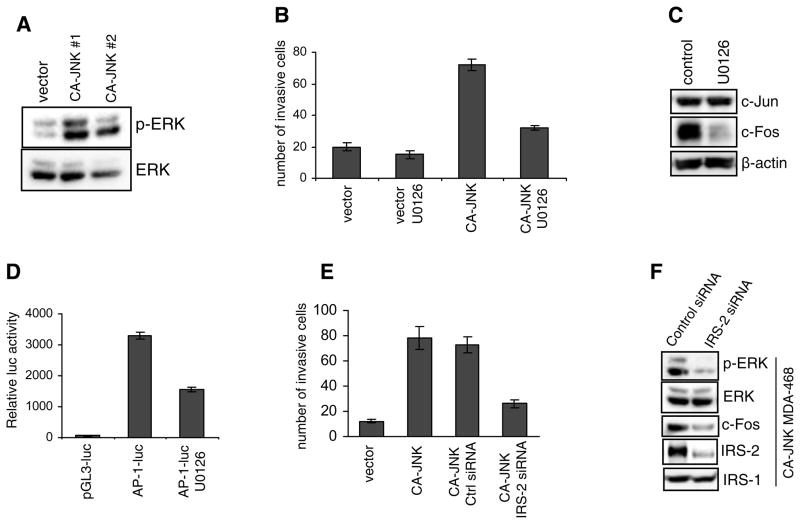
Figure 4.
Sustained JNK activates ERK. (A) Immunoblotting of total and phosphorylated levels of ERK in control and CA-JNK-overexpressing MDA-MB-468 cells. (B) The transwell invasion assay was conducted using control and CA-JNK cells. The inhibitor U0126 was used to block ERK activity. Each bar represents mean ± SD of samples measured in triplicate. (C) Expression of c-Jun and c-Fos was examined in CA-JNK-expressing cells treated with U0126 for 24 h. (D) CA-JNK-expressing cells were transfected with an AP-1-luc reporter construct and then treated with U0126 for 24 h, followed by luciferase assays. A β-galactosidase vector was used for normalization. (E) CA-JNK-expressing MDA-MB-468 cells were transfected with control siRNA or IRS-2 siRNA for 48 h. Cells were trypsinized and subjected to the transwell invasion assay. (F) CA-JNK-expressing cells were transfected with control or IRS-2 siRNA for 72 h. Levels of phosphorylated ERK and c-Fos were examined by immunoblotting. The IRS-2 homog IRS-1 was used as a control for confirming IRS-2 siRNA specificity.

