
Fig. 4.
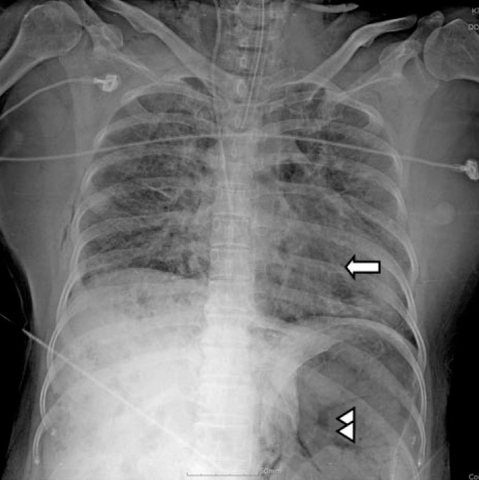
Chest X-ray taken at the scene revealing air density occupied in heart chambers (arrow) and hepatic vasculatures and splenic vein (arrow heads) indicating a massive air embolism.
Official websites use .gov
A
.gov website belongs to an official
government organization in the United States.
Secure .gov websites use HTTPS
A lock (
) or https:// means you've safely
connected to the .gov website. Share sensitive
information only on official, secure websites.
Chest X-ray taken at the scene revealing air density occupied in heart chambers (arrow) and hepatic vasculatures and splenic vein (arrow heads) indicating a massive air embolism.