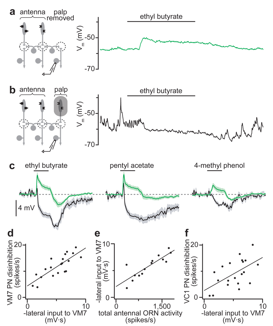
Figure 2. Lateral inhibition suppresses spontaneous EPSCs and scales with total ORN input.
a, Left: experimental configuration. Right: recording from a PN in glomerulus VM7. Olfactory stimulation evokes depolarization. Spontaneous EPSPs are absent.
b, Left: palps are shielded from odors. Right: recording from a PN in glomerulus VM7. Olfactory stimulation suppresses spontaneous EPSPs.
c, Average VM7 PN responses as in a (green) or b (black), n = 6–7 PNs for each condition. When ORNs are shielded (black), inhibition dominates. When ORNs are absent (green), excitation dominates throughout the stimulus period. (Off-inhibition likely reflects lateral postsynaptic inhibition, see Supplementary Fig. 5.)
d, Lateral input to VM7 (as in b) is correlated with the disinhibition in VM7 PNs after antennal removal (see Fig. 1b). Each point represents a different odor. Lateral input is measured as the time-integrated change in membrane potential.
e, Lateral input to VM7 is correlated with total antennal ORN spiking activity evoked by each odor.
f, Lateral input to VM7 is correlated with the disinhibition in VC1 PNs after antennal removal.

