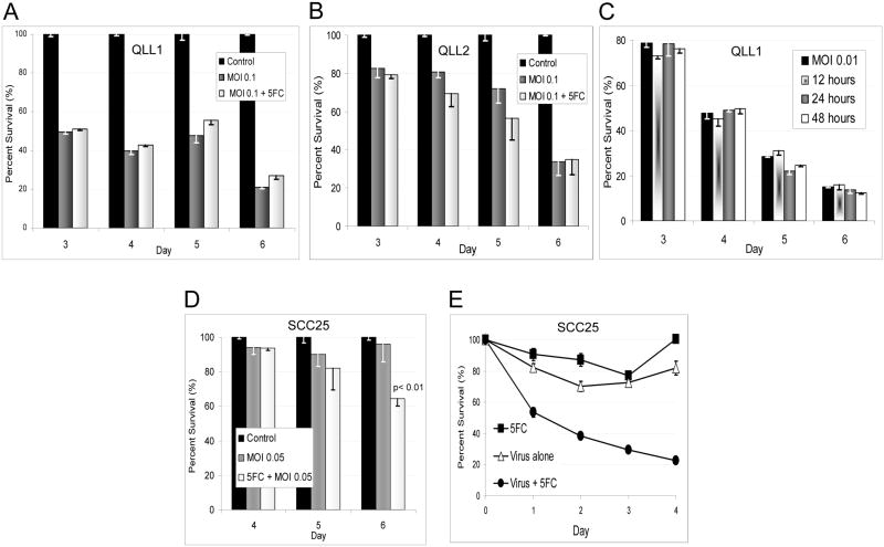
Figure 4.
A. The addition of the prodrug 5-FC at 600 μmol/L to QLL1 cells infected with OncoVEXGALV/CD at MOI 0.1 failed to enhance cytotoxicity as compared to cells that did not receive 5-FC. B. QLL2 cells treated identically demonstrated a subtle trend towards increased cytotoxicity with the 5-FC at days 4 and 5, but this difference was not statistically significant. C. To account for the possibility that 5-FU production interfered with viral replication, the timing of 5-FC addition to QLL1 at MOI 0.01 was varied and the 5-FC concentration increased to 2400 μmol/L. There were still no differences in cytotoxicity when the 5-FC is added at 12, 24, or 48 hours as compared to controls not receiving 5-FC. D. As direct viral oncolysis might limit the ability of infected cells to adequately express CD/UPRT prior to cell death, we treated the least sensitive SCC25 cell line with OncoVEXGALV/CD at MOI 0.05, with or without 5-FC at 2400 μmol/L. Under these conditions, direct viral oncolytic effects were minimal, but we observed a significant enhancement of cytotoxicity by day 6, with an increase from 4% to 35% cytotoxicity appreciated with the addition of the 5-FC (p<0.01). E. To assess the production of active 5-FU by OncoVEXGALV/CD, we quantified the cytotoxic activity of conditioned media from SCC25 cells exposed to OncoVEXGALV/CD and 5-FC, after viral inactivation. LDH assays on SCC25 cells demonstrated 80% cytotoxicity by day 4 for the cells treated with conditioned media collected from cells treated with both OncoVEXGALV/CD and 5-FC. There were significant differences at all time points (p≤ 0.01) in comparison with the 5-FC or virus only control samples. The slightly reduced viability detected from the 5-FC or virus alone samples likely results from growth rate reduction effects from nutrient-depleted media.

