
Fig. 6. The IFITM protein family restricts West Nile virus and dengue virus infections.
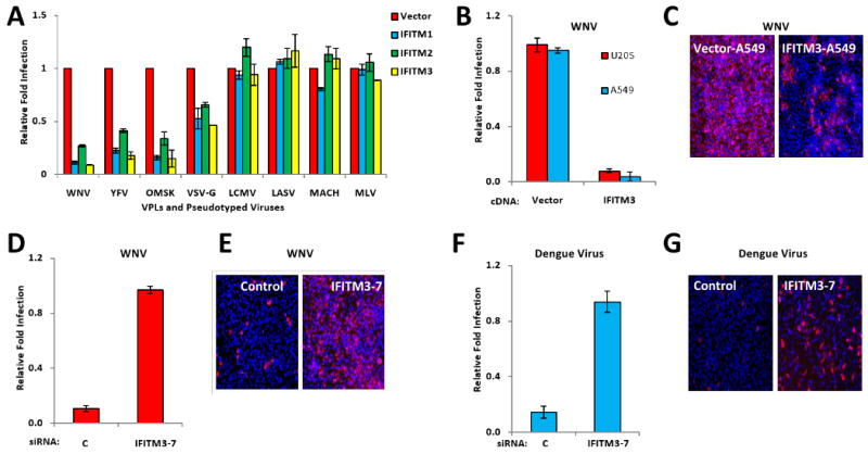
A) Vero E6 cells were transduced with retroviruses expressing the indicated IFITM proteins, or the empty viral vector. Two days later, the cells were incubated with flaviviral viral like particles (VLPs), expressing EGFP, and coated in envelope proteins from WNV, yellow fever virus (YFV) or Omsk virus (OMSK), or with EGFP-expressing MLV viruses pseudotyped with the indicated viral envelope proteins. Viral infection is expressed as mean EGFP fluorescence relative to vector control cells, as measured by flow cytometry 48 h post-infection. Values represent the mean ± SD, N=3.
B) A549 or U2OS cells stably expressing either IFITM3 protein or the vector alone (also shown in 4C, D), were infected with infectious WNV (strain 2741). 24 h. later, the cells were fixed and stained for viral E protein expression by IF. Values represent the mean ± SD, N=3
C) Images of A549 cells in B (red: WNV E protein, blue: nuclei), 4× magnification.
D) HeLa cells were transfected with the indicated siRNAs for 72 h, then infected with WNV. 24 h later, the cells were fixed and stained for viral E protein. Values represent the mean ± SD, N=3. C, Non-targeting siRNA negative control.
E) Images of HeLa cells in D (red: WNV E protein, blue: nuclei), 4× magnification.
F) HeLa cells were transfected with the indicated siRNAs for 72 h, then infected with dengue virus (New Guinea C strain). 30 h. post-infection, the cells were fixed and stained for viral E protein expression by IF. Values represent the mean ± SD, N=3. C, Non-targeting siRNA negative control.
G) Images of HeLa cells in F, 4×.